Allograft OATS® Technique
Precisely and reproducibly recover and reimplant hyaline cartilage.
.webp)
For large, symptomatic cartilage lesions, the allograft OATS technique combines precise instrumentation with the well-documented outcomes of cartilage restoration using fresh osteochondral allografts.
Arthrex is committed to providing fresh osteochondral allografts in a timely manner through its strategic partnerships with leading tissue banks focused on the highest standards for procurement, donor screening, and selection criteria. Tissue processing by our American Association of Tissue Banks (AATB)-certified partners balances safety with quality by ensuring the preservation of inherent biological properties of the tissue.
Everything needed for the allograft OATS technique.

Recovering allograft plugs from 15 mm to 35 mm is made easier with the allograft OATS instrument sets. Featuring a vice workstation and donor harvesting kits with depth stop reamers for exact graft/socket depth matching, achieving a press-fit graft does not require additional fixation implants.

Ideal for oblong lesions of the patella, the BioPatella™ OATS instrumentation features a series of precisely designed cutting instruments to overcome the challenge of the patella’s natural curvature through recovery of a graft that matches the articular surface. The BioPatella OATS instrumentation set is provided alongside the allograft OATS instrumentation, to address any patellar lesion type on the fly.

Specialized talus OATS® instrumentation facilitates harvesting 6 mm, 8 mm, 10 mm, 12 mm, and 15 mm osteochondral cores from an osteochondral allograft, providing a reliable outcome when restoring articular cartilage to the talus.1,2
Through a series of precise steps with dedicated instrumentation, replace damaged articular cartilage defects with healthy, fresh osteochondral tissue that matches the contour and cartilage morphology of the talus.
.webp)
Biologic Solutions
Arthrex biologic solutions, such as Arthrex ACP® platelet-rich plasma or AlloSync™ demineralized bone matrix, can be used to further support graft incorporation.
Take a Second Look
A 34-year-old male presented with persistent medial-sided right knee pain. For the past few months, the patient has experienced more intense swelling and stiffness, an uncomfortable new-onset “popping” sensation, and occasional catching. Complete arthroscopic joint evaluation showed the medial joint was being overloaded due to meniscal deficiency and varus alignment. The treatment plan included multiple joint preservation procedures, including allograft OATS® procedures of the medial femoral condyle (MFC) and trochlea, a medial meniscus allograft transplantation, and a tibial tubercle osteotomy.
2 Years Post-Op
Two years after these procedures, the patient was experiencing catching on the medial side of knee, and swelling with increased activity, and expressed interest in additional operative management with arthroscopy and evaluation of articular cartilage. After debridement and hardware removal, second-look images of the osteochondral allograft transplantation showed full graft incorporation.
Medial Femoral Condyle
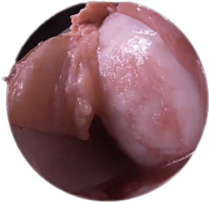


Trochlea


Courtesy of Brian J. Cole, MD, MBA and team
Reimbursement and Claims Guidance
Arthrex provides comprehensive reimbursement and claims support, including product-specific information about coding and billing scenarios that may be used for procedures that employ our products.
Note: Information contained in this reimbursement guide is for educational and strategic planning purposes only.
2 Years Post-Op
Two years after these procedures, the patient was experiencing catching on the medial side of knee, and swelling with increased activity, and expressed interest in additional operative management with arthroscopy and evaluation of articular cartilage. After debridement and hardware removal, second-look images of the osteochondral allograft transplantation showed full graft incorporation.
Courtesy of Brian J. Cole, MD, MBA and team
Medial Femoral Condyle
Trochlea

